PREGNANCY 0-14. WEEK
Genes and Chromosomes
The genetic information in sperm and egg is hidden in chromosomes. (23 chromosomes from the mother, 23 chromosomes from the father) Chromosomes each consist of tightly wound DNA sequences. When the DNA molecules in a human cell are dissolved and added end to end, its length can exceed 180 cm. 20,000-25,000 genes make up a human, about the same number it takes to make up a chicken. We all carry a set of genes arranged in 23 pairs of chromosomes in the nucleus of almost every cell in our body. Our genes are a series of commands that tell us whether we will be a fish or a tree, but a human being, and even determine what kind of person we will be. Our mother and father have equal shares in our genetic makeup, but it is the father who determines the gender of the child. The special function of the 23rd chromosome pair is to determine gender. The sex chromosome in the egg inherited from the mother is always the same type: type X. But in the sperm, the sex chromosome inherited from the father can be of two types: the X chromosome if it is a girl, and the Y chromosome, which is a much smaller chromosome, if it is a boy. The parents of this baby will be able to learn the gender of this baby months later, but if it is an X sperm that wins this fertilization race, it will be their daughter, and if it is a Y sperm, it will be their son. The genes this baby receives from its parents have already determined its appearance and, to a large extent, its character.
Everything is in this mix...
In humans, all body cells, except the nucleated mature red blood cells, have a total of 46 chromosomes, 23 from the mother and 23 from the father. When sperm and eggs are formed, this number is divided in half by a special type of cell division. During this division, the chromosomes remain in their respective pairs. Then, the parts forming each pair are collected at opposite poles of the dividing cell. Thus, sperm and egg cells contain genetic combinations that are different from each other and from the cells of their parents. While fertilization ensures constant diversity in the human species, the number of chromosomes is always maintained at 46.
The journey begins... Day 2
Immediately after fertilization, the egg travels through the fallopian tubes to the uterus. After about 30 hours, it starts to split first thing. Since each cell in the body requires a copy of the original genetic message for that cell, the chromosomes first multiply and create an identical copy of their own DNA. These original and duplicate chromosomes are collected and arranged in the middle of the egg on a specially formed spindle structure. The threadlike extensions of this spindle structure shorten and pull the chromosomes towards opposite poles of the nucleus. When the chromosomes separate into two sets, the nucleus divides in two and the cell is divided into two identical, smaller cells containing exactly the same genetic information. While sex cells (sperm and eggs) are produced in the testes and ovaries, primary sex cells reduce their chromosome number to half the chromosome number of other cells. This requires two special cell divisions. First, the primary germ cell divides into two cells, each with a set of chromosomes. Both of these cells copy their chromosome sets and divide again. Thus, one set of chromosomes falls into each of the four resulting cells. This process continues in men, starting from puberty; four sperm are produced from each primary sex cell. In females, it begins before birth, when the fetus is three months old, but the first round of cell division is not initiated until puberty.
Cell ball Day 5
The division process continues as tiny clusters of cells make their way into the fallopian tubes. Since the proliferating cells depend on the nutrients stored in the egg before fertilization, they become smaller with each division, so the entire cell cluster can never be larger than the original egg and remains completely covered by the zona pellucida. At the eight-cell stage, the cells change shape, come together, and become compacted into a compact sphere. Mulberry andiron This sphere with 12-15 cells is called morula. When it reaches the uterus three to four days after fertilization, it has 32-64 cells. Over the next two to three days, it becomes a sphere called a blastocyst, filled with nutritious fluid from the inner uterus. When the blastocyst grows, it divides into two parts. The outer layer of the flattened cells, called trophoblast, will become the placenta in the future, and the cluster of cells inside the sphere will become the embryo. These inner cells are known as embryonic stem cells. These cells have an extraordinary ability. They can develop into 350 different cells and become any part of the body. Early stage cells have greater transformation potential.
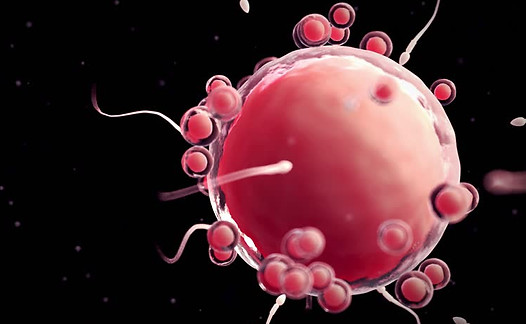
Day 1 of Pregnancy
The sperm now faces the daunting task of passing through the follicle cells around the egg and penetrating the egg's jelly-like covering. It has special equipment for this. It has a cap filled with biological enzymes on its head. Working like a chemical drill, these enzymes discharge through the holes in the cap, distribute to the follicle cells and enable the sperm to penetrate the zona pellucida. No single sperm can overcome this obstacle on its own, without the help of hundreds of other sperm. As soon as the sperm touches the zona pellucida, it locks onto this target surface and a complete transformation occurs. Under the pressure of its revived tail, it secretes other enzymes that break down the path from the zona pellucida to the egg membrane. The sperm is now naked; His headgear has disappeared and the membrane on his head has broken down and dispersed. There is no prize to be given to the runner-up. When the fast-acting sperm penetrates the egg membrane, a change occurs in the egg and the zona pellucida, making it impossible for another sperm to pass through. The head and tail of the winning sperm enter the egg, leaving behind the sperm membrane. The tail is separated from the head and the nucleus of the sperm is pulled towards the nucleus of the egg. These two nuclei become a single nucleus. Fertilization is completed. Two separate genetic sequences, one from the mother and one from the father, combine to create a new genetic code. The moment when a person becomes his own, something that has never existed before and will never be repeated, is the moment he falls into the womb.

Implantasyon 2.Hafta
Yedinci güne gelindiğinde, artık yaklaşık yüz hücre içeren blastosist, progesteronun etkisiyle yumuşayan ve kabaran rahim içi duvarına gömülür. Rahim içi duvarına yapışan, zarlarını kaybederek bir araya gelip kaynaşan trofoblast hücrelerinden aldığı destekle blastosist hızlı ve saldırgan bir şekilde rahim içi duvarına yayılır. Rahim hücrelerine kendilerini tahrip etmelerini söyleyen sinyaller veren moleküller ve bu hücrelerden geriye kalanları sindiren enzimler üreterek yerleştiği yerdeki rahim hücrelerini oyarlar. Aynı zamanda, trofoblast, süngerimsi çıkıntılar oluşturarak rahim duvarının derininde annenin kan dolaşımından yol açarlar..Annenin kanının, oluşan bu deliklerden dolmasıyla plesantada bir kan rezervi oluşur, ilk emriyo hücreleri buradan beslenir ve hayatta kalır. Onuncu güne gelindiğinde, hala mikroskobik büyüklükteki embriyo rahim içi duvarına gömülerek kaybolur, geride bıraktığı yara izi iyileşmeye başlar, bu nedenle dokusu embriyoyu geçici bir süre koruyan bir kapsül görevi görür.
Organizasyon 3.Hafta
Rahim duvarında kendine bir yuva kurduktan sonra embriyonun artık kendini organize etmesi gerekir. Yaklaşık 13. günde, iç hücre kitlesi, biri diğerinin içinde iki kabarcık oluşturur. Bunlardan biri, ilk ektodermdir (dış deri), doğrudan trofoblastın iç yüzeyine bağlıdır ve ileride amniyon boşluğu haline gelecektir. Embriyo iki kabarcıklı diskte şekillenmeye başlar. Gastrülasyon adı verilen bu hareketli süreçte hücreler topluca hareket eder. Üçüncü haftanın başında, embriyo diskinin ortasına doğru boyuna bir oluk açılmaya başlar. Hücreler, bu oluğa doğru hareket eder
And they throw themselves into it. The result is an embryo consisting of three initial layers. These layers will later become the organs of the body. The upper, ectoderm layer, along with the eye lenses and most of the nervous system, will become the outer layers of skin, hair, nails, nipples, and tooth enamel. Once the cells in the initial layers are directed or programmed for a certain function, it is no longer possible for them to change and become another cell type.
Heads or tails
How does a human with billions of cells emerge from a newly fertilized egg thousands of times smaller than a centimeter? How do cells know that they are inside the embryo and which tissue or organ they belong to? In the end, how does one create a perfect baby with everything in place from head to toe?
The cells, which are completely identical at first, somehow specialize and turn into one of the 350 special types of cells found in the brain, bone, muscle, liver or in mammals like us. In genetic disorders and cancer, something may go wrong in the control of genes. In healthy cells, the button for suitable genes is pressed, the button for unsuitable genes is turned off, and thus, each plays its part in the formation of the embryo. When the fertilized egg grows into an embryo, it is initially a small, shapeless mass. Gradually two asymmetries are formed; a head-tail axis and a face-dorsal axis.
Brain and Spinal Cord Week 3
Since the nervous system coordinates the activities of other systems of the body, it begins to form very early and continues to develop until birth and even after birth. By the end of the first month, the embryo will have laid the foundation of the entire nervous system. The primitive neurons of the nervous system will come together to form nerves and eventually grow and extend to the smallest point of the developing body, sending and receiving messages at a speed of 480 km per hour. On day 18 or 19, when the embryo is only 1.5 mm long, the spinal cord initiates a transformation in the ectoderm. Ectoderm cells thicken and begin to collapse inward to form a scar extending along the back of the embryo and form a flat layer. The two slopes of the scar curl towards each other and close on each other on the 23rd day, creating a hollow, empty canal. This canal is called the neural tube. is given. As the nerve fibers form, the foundations of other organs begin to be laid. Tissue blocks like stacks of bricks begin to appear on both sides of the incipient nerve fiber, initially a few, then 10, 20 and 40. Soon, various parts of the nervous system begin to form from different parts of the neural tube. Three large lobes appear before the tube unites. By the end of the third week, the three main parts of the brain have formed, namely the hindbrain, midbrain and forebrain. The spinal part of the tube is the part that changes the least; even in our adult lives, the spinal cord is still a thick-walled tube with a canal running through the middle. If the neural tube cannot close at the end towards the head, then the brain does not form, this condition is called anencephaly. Children born with this abnormality are usually stillborn or die within a few days after birth.
Signs of Life
Five weeks have passed since the start of the mother's last menstrual period. Failure to have menstrual bleeding when it should be is usually the first sign that the mother may be pregnant. In the next five weeks, mothers may notice incredible things happening in their bodies and realize that they are pregnant without needing a test. By the third month, there is a significant increase in the amount of blood pumped by the mother's heart. This amount increases as pregnancy progresses and increases by 30-40 percent until birth. This increase is mainly due to the heart beating harder, but its speed also increases. After the embryo settles into the mother's womb, it receives all its needs for growth from the mother's bloodstream. Moreover, they are ready to provide the baby with the necessary nutrients after birth. The mother's breasts grow and the veins on their surface become more and more prominent. The gland tissue maturing under the influence of changes in hormone levels causes sensitivity and tingling in the breasts. Although the hormones that cause milk secretion are also produced during pregnancy, milk secretion is suppressed until birth. However, in the last trimester of pregnancy, a clear fluid called colostrum may begin to leak from the nipple. Colostrum is a cocktail of sugars, proteins, and antibodies. It meets all the nutritional needs of the baby and protects the baby against infections in the first few days after birth until real milk starts to come. Other changes that occur under the influence of hormones are a loosening of the joints that facilitates labor and birth and the formation of brown spots on the skin. Birth stretch marks appear due to the effect of the collagen under the skin, which stretches and cracks to adapt to the mother's expanding body. Increased blood flow to the kidneys increases urine production andIt causes the candidate to go to the toilet more frequently. The mother's appetite usually increases. However, the extra energy she needs throughout the entire pregnancy is at most 60,000 calories. Although the amount of nutrients the mother needs does not increase much, attention should be paid to the type of food. He may need especially iron and vitamin supplements. The most visible sign of pregnancy is the growth of the mother's belly. The uterus, trying to adapt to the growing fetus, expands greatly. It begins to appear from the groin at approximately the 12th week, rises to the navel at the 22nd week, and rises to the ribs at the 36th week. This stretching will not worry the mother at all. Under the influence of pregnancy hormones, the smooth muscles in the mother's uterus and abdomen grow and stretch, increasing 60 times during pregnancy, but almost returning to their previous state 6 weeks after birth. Biologically, pregnancy is a normal situation for a woman of reproductive age. This view frees us from the view that pregnancy is a kind of disease and allows us to see the unpleasant symptoms that occur with pregnancy as an inability to adapt to the hormonal and physical changes that occur due to the growing fetus.
Fracture Week 4
The embryo has now begun to take on a slightly human form. Moreover, the embryo has also formed the other most important membranes of its own life support system. The endoderm bubble remains in the abdomen of the embryo like a balloon suspended from its navel. This is the yolk sac. Even though there is no yolk in the egg in humans, the embryo meets all the nutrients it initially needs from this sac until the baby's placenta is fully developed. The yolk sac also forms part of the digestive tract and produces primitive blood cells and germ cells. As the embryo rolls over, the ectoderm bubble stretches and becomes a balloon that almost completely surrounds the embryo. This balloon is called amniotic sac. It is filled with fluid in which the fetus floats during the time it spends in the womb. The yolk and amniotic sacs are suspended by a ligament stalk from a larger balloon called the chorionic sac.
A heart begins to beat... Week 4
The cardiovascular system is one of the first important systems to start working in the embryo. Without the heart and vessels, it is not possible to deliver the nutrients and oxygen required for the growth and development of the embryo. The nutrients that the embryo initially received from the egg and yolk sac have already been exhausted. At the beginning of the third week, blood vessels begin to appear, first in the yolk sac, ligament stalk and allontois, and two days later in the embryo itself. These vessels consist of hollow and stacked clusters of mesoderm cells. At this stage, the bulk heart cells are still quiescent. This mass of cells, initially arranged like a double crescent-shaped pipe, fuses together and turns into a single 'S' shaped pipe. At the beginning of the fourth week, on the 22nd or 23rd day, a single heart cell is suddenly killed by a jolt. By the end of the fourth week, along with the rhythmically beating heart, primitive blood cells begin to circulate in the hair-thin vessels inside the fetus.

Bodybuilding Week 4
On the 28th day, the embryo, which is growing approximately 1 mm per day, is still the size of the head of a matchstick, but its vital organs are already evident. The buds that appear on both sides of the body will become its arms and legs in the future. Its miniature heart protruding from its body beats 80 times per minute and gets faster every day. At the end where the head is located, five skin folds like gills are visible, these folds will form the face and jaw. Although it is not yet fully functional, the tube-shaped digestive system descends from the mouth that has now opened. There is a stomach, a liver, a gallbladder, a pancreas and a thyroid gland. For the next four weeks, our embryo will continue to grow, following its own internal genetic commands. All organs and systems will begin to take shape and the baby will have a human appearance.
Head 6th week
The embryo is 10-12 mm long in the 6th week of its development. Arm buds are wing-like, and leg buds are fin-like. The cell layers that will form the hands and feet appear. A few days ago, his eyes, which consisted of black dots, appeared. Their retinas have some pigment, they have lenses, and their eye muscles are starting to form. The nose and mouth of the embryo can be selected. In the sixth week, the embryo's brain consists of 5 cavities lined with embryonic nerve tissue that will completely fill it in the future. The foundations of the cerebral cortex have been laid. Kidneys have formed, but they do not purify waste. The lobes of the lungs have appeared. The shape of the external reproductive organs appears as a bulge. However, so far the male and female embryos look exactly the same.
Human Face Week 7 and 8
In the past 5 weeks, the embryo has formed hundreds of different types of cells, its muscles and nerves are twitching, and its major organs and systems are all complete. The embryo now has distinctive human features, but its head is still disproportionately large for its body, measuring half the head-to-rump distance. His hands and feet stand close together in front of his body. Light-sensitive cells of the retina have formed and nerve connections have been established from the retina to the brain. It will not open again until 25-26 weeks, until the nerves of the visual center in the cerebral cortex are ready. There is a tiny tongue inside its mouth, where the palate and taste buds have begun to form. Consider the 20 milks in the developing jawbone, all of them have ready tooth buds. The intestines, which are separated from the heart and lungs by the diaphragm, grow so rapidly that the intestinal loops formed pop out of the abdominal wall of the embryo for a while and continue to grow in a sac close to the umbilical cord.
Planned Cell Death
As the baby's legs take shape and lengthen, the skin layers that form the legs begin to thicken and form the cartilage that will become hard bone in the future. This cartilage only acts as a mold. Bone cells flock to this cartilage and replace it. At the end of the eighth week, the webs on the embryo's hands and feet lift, the fingers separate, and shoulders and elbows appear. The baby gets rid of unnecessary tissues in his hands and feet and forms fingers and toes. The brain is shaped in a similar but more subtle way, by creating more cells than necessary and destroying cells that do not fit the pattern of interconnectivity properly. Cell suicide appears to be the result of a developmental compromise. Planned cell death serves to limit cells within an organ or tissue to a certain number. Blood and skin cells are constantly renewed by progenitor (source) cells, but this proliferation must be balanced by cell death. In an adult, 50-70 billion cells die every day due to programmed cell death. The sum of the cells that proliferate and are subsequently destroyed in a year is equal to the weight of a human being.
From Embryo to Fetus
After eight weeks of growth, the embryo (embryo, which means “growing” in Greek, becomes a fetus, which means “son” in Latin). First, most of the embryo's vital internal and external structures had survived the most critical period of development. Secondly, by the second month, the yolk sac becomes redundant and shrinks and disappears. The placenta undertakes this extremely important task of feeding the fetus. The placena, which provides nutrients and oxygen to the fetus, also filters carbon dioxide and other waste materials.

Placenta
Placenta is a Latin word meaning flat round mold; It resembles a spongy, pizza-shaped liver. The placenta is adorned with extraordinarily fine blood vessels that surround the chorionic villus network, most of which remains within the mother's blood reserve. This reserve fills the space formed between the spongy protrusions of the trophoblast, which clings tightly to the intrauterine wall. Chorionic villi, which protrude into the intervillous space, are the newest projections that will replace the trophoblast cells of the chorion. Just as a tree is nourished by its roots from the soil, the blood vessels of the chorionic villi absorb everything the fetus needs from the mother's blood. At the end of the third week, the embryo's blood begins to flow through the small vessels in the chorionic villi, and after a week, the mother's blood begins to penetrate into the intervillous space. The placenta grows faster than the fetus in the first three months of pregnancy. It reaches the same weight as the fetus in the 15th week. At birth, it is one-sixth of the weight of the fetus. Any bleeding that occurs during pregnancy is from the mother's blood, not the fetus's. Even if the placenta is damaged, the circulation of the fetus is protected.
Chorionic Villus Test
Chorionic villus test or CVS test is a prenatal diagnostic method that helps detect genetic abnormalities in the fetus. Generally, it is around the 11th-13th week of pregnancy. weeks, a small cell sample is taken from the chorionic villi. With this test, Down syndrome, cystic fibrosis, Tay-Sachs disease, sickle cell anemia and
Diseases such as thalassemia are detected. About 1% of women have a miscarriage after CVS. The risk of miscarriage after CVS is slightly higher than the risk of miscarriage after amniocentesis. The reason may be that CVS is performed at an earlier stage, when the fetus is already likely to miscarry.
Illegal Fetal Cells
When it first forms, the placental membrane is a single cell thick, and the fetus's blood cells sometimes leak through microscopic gaps and enter the mother's circulation. There may be as many as 1 in 100,000 fetal blood cells in the mother's blood, and this rate is higher in fetuses with chromosomal abnormalities. These cells can remain in the mother's bloodstream for decades after the baby is born. Researchers have found that male fetal stem cells survive in the mother's bone marrow for more than 50 years after birth. This may also explain why bone marrow transplanted from a mother to a child is less likely to be rejected than bone marrow transplanted from a father to a child. The fetal cells remaining in the mother can ensure that the bone marrow transplanted from the mother is not perceived as foreign by the child's body.
Umbilical Cord
The placenta provides the embryo with everything it needs, carries its waste and protects it from harmful unwanted guests. It does all of this with the baby's lifeline, the umbilical cord. When the embryo first implanted in the uterine wall, it was small enough to exchange enough gas, nutrients, and waste. Later, in the early stages of growth, as the uterus rearranges according to the embryo, a short stalk is formed from its own tissue. As the embryo's heart and circulation develop, the blood vessel rings inside the stalk also grow. As the fetus grows in its amniotic sac, the stalk lengthens and the blood vessels become part of the placenta, which is growing at its tip in the uterus. When the 40th week of pregnancy is over, the umbilical cord, which is approximately 55 cm long, is coiled like a hose within the amniotic sac, allowing the mature fetus to move freely. Since the veins are longer than the cord itself, it is common for the umbilical veins to twist and turn. They usually take the shape of a ring, forming pseudoknots that are not harmful to the baby. However, in one in 100 pregnancies, a knot actually forms in the cord. The knot may tighten and cause the fetus to have difficulty breathing. Usually, during birth, a knot is formed as a result of the fetus passing through the ring of the cord. Since these knots are usually loose, they do not cause concern. In about one-fifth of safe births, the cord forms a loose loop around the fetus's neck.
Growth Surge 9-12. Week
The fetus's body quickly tries to catch up with its head, and the crown-rump distance doubles within four weeks. As its pointed tail retracts into its body, it loses its previous comma-like shape. As his back straightens and straightens, his neck is exposed and he lifts his chin from his chest. His face lacks detail, with eyes set far apart on either side, lips stuck together, and ears drooping. Buds of permanent teeth begin to form under the gums. Towards the end of the 12th week, the first quince-like hairs begin to appear. His nails start to grow. Her arms approach their final proportions, yet her legs are still short, her thighs noticeably small. His intestines, which are pulled back from the umbilical cord towards his abdomen, now protrude less. In the ninth week, the primary site of formation of red blood cells is the liver, while in the 12th week, red blood cell production activity in the liver slows down and begins to accelerate in the spleen. The kidneys begin to produce urine and the gallbladder begins to secrete bile. Additionally, it is possible to detect heartbeats with electronic devices starting from the 10th week.
Urine Production
By the 20th week, the fetus produces two to three teaspoons of urine per day. Although it follows a very circuitous route before completely excreting urine, it continues to excrete this urine throughout the time it spends in the womb. When his bladder fills and empties every 20-30 minutes, his urine is thrown into the amniotic fluid he swallows. This swallowed waste is reabsorbed through the digestive tract, into the bloodstream of the fetus, and then finally passed into the placenta.
Hairs
9-12. The hairs that start to form in weeks are not noticeable until the 23rd week. First, the eyebrows, upper lips and chin grow hair. Each hair grows from a hair follicle in the lower layer of the skin.

The body moves...
In the third month, the fetus begins to move. The nervous system develops rapidly, producing an average of 2.5 million neurons per minute. His whole body starts to move. Over the next 4 weeks we can see him kicking, turning his feet, and curling his toes. He will bend his elbow and wrist, make a fist with his little hands and cover his face with his hands. He will squint his tightly closed eyes, frown, purse his lips and open his mouth. He will begin to react to touches. These movements play an extremely important role in the development and growth of muscles and strengthening of joints. Because the fetus is too small for the mother to feel these movements yet. The brain cannot yet control the movements of the fetus at this stage. These movements are involuntary reflex type contractions.
Introspection
A pregnant woman is 10-14 days old. may undergo an ultrasound scan per week. The use of ultrasound to look inside the womb has radically changed our knowledge and understanding of fetal development and maternal care. Looking at ultrasound does not pose any danger, unlike x-rays. Ultrasound scans do not cause any harm to the fetus. In the womb, the fetus lives in fluid. Even their lungs are filled with fluid, and ultrasound provides moving images that tell a lot about fetal health and development to anyone who knows how to look. With this screening, it is possible to detect early, distinctive diagnostic features of genetic abnormalities. For example, an obstetrician who evaluates measurements of the part of the fetal neck in fluid, called a nuchal fold scan, together with measurements of two substances in the mother's blood, can strengthen the detection of Down syndrome. This method does not definitively say that the fetus has Down syndrome, but it allows a better calculation of the risk. Estimating the date of birth based on the size of the fetus during the first scan to check the baby's health may be more accurate than the estimate based on the date of the mother's last menstrual bleeding. Additionally, ultrasound determines whether the mother is pregnant with one fetus or two or more fetuses.
Amniotic Fluid
Amniotic fluid is of vital importance in the growth and development of the fetus. Amniotic fluid, which consists largely of the fetus's urine, placenta and tissue fluids from the fetal lungs, is constantly reabsorbed and renewed in such a way that its fluid content changes completely every 3 hours. Some of the amniotic fluid passes through the membranes of the amniotic and chorionic sacs into the blood vessels of the uterus, and some is absorbed by the developing lungs and digestive tract of the fetus through the umbilical cord. The volume of amniotic fluid is approximately 25 ml at the 12th week. It increases rapidly from the middle of pregnancy, reaching a peak of 0.9 liters at the 34th week, but then mysteriously drops to 280 ml at the 42nd week.
amniocentesis
Amniocentesis is a prenatal examination method performed by taking a sample of amniotic fluid from the amniotic sac. The cells, proteins, minerals and other chemicals in the amniotic fluid tell the physician a lot about the health and development of the fetus. It is also possible to collect amniotic fluid from the cervix towards the end of pregnancy. However, amniocentesis should still be performed early in pregnancy, at the 15th or 16th weeks. During this period, a sample is taken by entering the abdominal wall with a long, thin needle. If there is a family history of the parents that suggests that the baby may have a serious genetic or chromosomal disease such as Down syndrome, amniocentesis may be required. Since pregnant women over the age of 35 are likely to be pregnant with a baby with a chromosomal abnormality, these women are often advised to consider amniocentesis. Preliminary tests of the mother's blood can sometimes support this possibility. Amniocentesis carries a slight risk of causing miscarriage.